HUMAN EYES
|
The human eye is an organ that reacts to light and has several purposes. As a sense organ, the mammalian eye allows vision. Rod and cone cells in the retina allow conscious light perception and vision including color differentiation and the perception of depth. The human eye can distinguish about 10 million colors.
Similar to the eyes of other mammals, the human eye's non-image-forming photosensitive ganglion cells in the retina receive light signals which affect adjustment of the size of the pupil, regulation and suppression of the hormone melatonin and entrainment of the body clock. |
|
EYE STRUCTURE
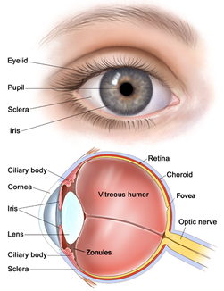
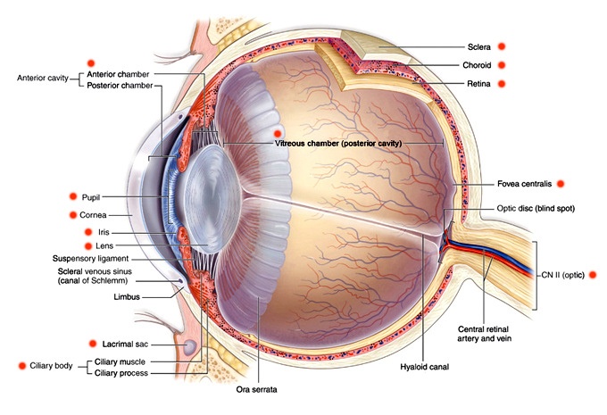
The eye is not shaped like a perfect sphere, rather it is a fused two-piece unit. The smaller frontal unit, transparent and more curved, called the cornea is linked to the larger white unit called the sclera. The corneal segment is typically about 8 mm (0.3 in) in radius. The sclerotic chamber constitutes the remaining five-sixths; its radius is typically about 12 mm. The cornea and sclera are connected by a ring called the limbus. The iris is the colored circular structure concentrically surrounding the center of the eye, the pupil, which appears to be black. The size of the pupil, which controls the amount of light entering the eye, is adjusted by the iris's dilator and sphincter muscles. A device known as an ophthalmoscope is used to see inside the eye.
Light enters the eye through the cornea, then the pupil and then through the lens controlled by ciliary muscles. Light falling on the light-sensitive cells of the retina is converted into electrical signals that are carried to the brain by the optic nerves.
Light enters the eye through the cornea, then the pupil and then through the lens controlled by ciliary muscles. Light falling on the light-sensitive cells of the retina is converted into electrical signals that are carried to the brain by the optic nerves.
EYE COMPONENTS
The eye is made up of three coats, enclosing three transparent structures. The outermost layer, known as the fibrous tunic, is composed of the cornea and sclera. The middle layer, known as the vascular tunic or uvea, consists of the choroid, ciliary body, and iris. The innermost is the retina, which gets its circulation from the vessels of the choroid as well as the retinal vessels, which can be seen in an ophthalmoscope.
Within these coats are the aqueous humour, the vitreous body, and the flexible lens. The aqueous humour is a clear fluid that is contained in two areas: the anterior chamber between the cornea and the iris, and the posterior chamber between the iris and the lens. The lens is suspended to the ciliary body by the suspensory ligament (Zonule of Zinn), made up of fine transparent fibers. The vitreous body is a clear jelly that is much larger than the aqueous humour present behind the lens, and the rest is bordered by the sclera, zonule, and lens. They are connected via the pupil.[5]
Within these coats are the aqueous humour, the vitreous body, and the flexible lens. The aqueous humour is a clear fluid that is contained in two areas: the anterior chamber between the cornea and the iris, and the posterior chamber between the iris and the lens. The lens is suspended to the ciliary body by the suspensory ligament (Zonule of Zinn), made up of fine transparent fibers. The vitreous body is a clear jelly that is much larger than the aqueous humour present behind the lens, and the rest is bordered by the sclera, zonule, and lens. They are connected via the pupil.[5]
HOW DO THE HUMAN EYES WORK?
With a number of ways, the human eye works much like a digital camera:
- Light is focused primarily by the cornea — the clear front surface of the eye, which acts like a camera lens.
- The iris of the eye functions like the diaphragm of a camera, controlling the amount of light reaching the back of the eye by automatically adjusting the size of the pupil (aperture).
- The eye's crystalline lens is located directly behind the pupil and further focuses light. Through a process called accommodation, this lens helps the eye automatically focus on near and approaching objects, like an autofocus camera lens.
- Light focused by the cornea and crystalline lens (and limited by the iris and pupil) then reaches the retina — the light-sensitive inner lining of the back of the eye. The retina acts like an electronic image sensor of a digital camera, converting optical images into electronic signals. The optic nerve then transmits these signals to the visual cortex — the part of the brain that controls our sense of sight.
